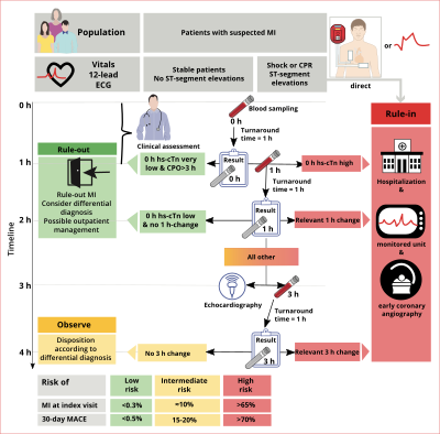
Myocardial Infarction Is Best Identified in Which Leads
Log in for more information. Prolonged deprivation of oxygen supply to the myocardium can lead to myocardial cell death and necrosis2.
Ecg Localization Of Myocardial Infarction Ischemia And Coronary Artery Occlusion Culprit Ecg Echo
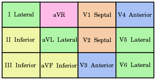
A posterior wall myocardial infarction is best identified in which leads.

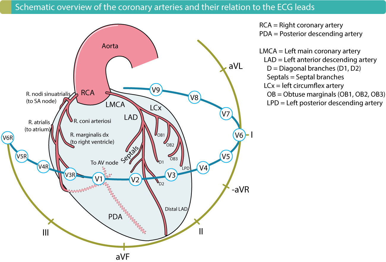
. With coronary artery occlusion the myocardium is deprived of oxygen. The right precordial leads V1 V2 and V3 are critically important in diagnosing posterior wall STEMIs. In other words acute myocardial injury acute myocardial ischemia acute myocardial infarction.
Most myocardial infarctions are due to underlying coronary artery disease the leading cause of death in the United States. A posterior wall myocardial infarction is best identified in which leads. Log in for more information.
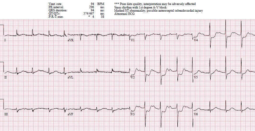
Automated interpretation of ECG is widely used as decision support for less experienced physicians. This occurs because these ECG leads will see the MI backwards. Log in for more information.
V1 and V2 C. Start studying 12-Leads and Myocardial Infarction. 12-Lead ECGs and Myocardial Infarction.
Although 80-lead ECG body surface mapping is more sensitive for ST-elevation myocardial infarction STEMI than the 12-lead ECG its clinical utility in chest pain in the emergency department ED has not been studied. An MI is often associated with atherosclerosis of the _____ arteries. A posterior wall myocardial infarction is best identified in V1 and V2.
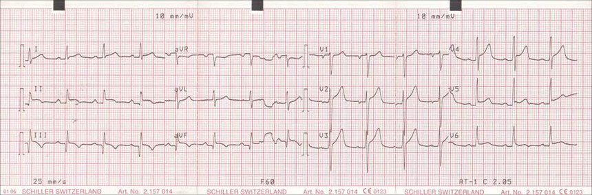
A posterior wall myocardial infarction is best identified in V1 and V2. The 12-lead ECG together with patient history and clinical findings remains the most important method for early diagnosis of acute myocardial infarction. A RS wave ratio greater than 1 in leads V1 or V2.
A posterior wall myocardial infarction is best identified in which leads. A posterior wall myocardial infarction is best identified in V1 and V2. A posterior wall myocardial infarction is best identified in V1 and V2.
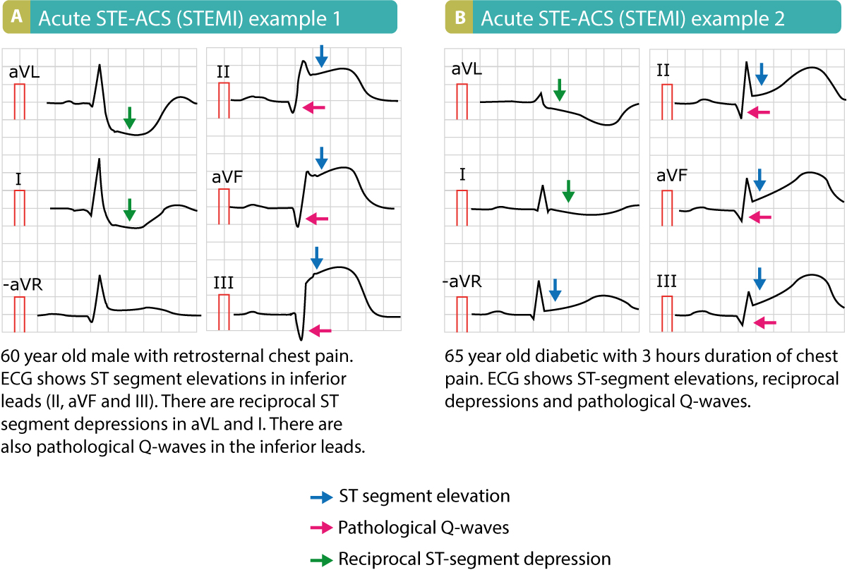
An ECG performed with the use of posterior leads revealed ST-segment elevation in leads V 7 V 8 and V 9 which was consistent with posterior-wall myocardial infarction. Knowing the site of infarction is essential in anticipating clinical manifestations and therefore has implications for the patients plan of care. Added 34 days ago782021 75823 PM.
Added 152 days ago782021 75823 PM. A posterior wall myocardial infarction is best identified in V1 and V2. Log in for more information.
Terms in this set 43 A _____ is a death of the heart muscle due to inadequate blood supply. Features measured on leads originating from the upper left precordial area lower midthoracic region and the back correctly identified 97 of anterior MI patients with a specificity of 95. None Where is the damage.
Learn vocabulary terms and more with flashcards games and other study tools. Log in for more information. Acute posterior wall STEMI should be suspected whenever the ST-segments are depressed in leads V1 V2 and V3.
We sought to determine the prevalence clinical care patterns and clinical outcomes of patients with STEMI identified on 80-lead but not on 12. Log in for more information. This blood exerts pressure on the heart.
Further details regarding evidence of acute myocardial ischemia elevated troponin levels myocardial injury and ischemic vs. Acute myocardial infarction and the 12-lead ECG. A posterior wall myocardial infarction is best identified in V1 and V2.
Log in for more information. V4R V5R V6R Artery. An acute myocardial infarction occurs when acute myocardial ischemia causes myocardial injury.
The leads are placed anteriorly but the myocardial injury is posterior. Locating Myocardial Damage Leads. A posterior wall myocardial infarction is best identified in V1 and V2.
A posterior wall myocardial infarction is best identified in which leads. Preload-amount of blood that is brought back to the heart the be pumped throughout the body. Myocardial infarction is typically the result of a blockage in one of the coronary arteries due to an _____.
Recent reports have demonstrated that artificial neural networks can be used to improve selected aspects of. It is important that critical care nurses are able to identify the location of cardiac muscle ischemia injury and infarction on the electrocardiogram. A posterior wall myocardial infarction is best identified in V1 and V2.
Ad Visit The Official Physician Site For An Acute Myocardial Infarction Treatment Option. A posterior wall myocardial infarction is best identified in which leads. Log in for more information.
Log in for more information. Together with patient history and clinical findings the 12-lead ECG is still the most readily available and best method for the early diagnosis of acute myocardial infarction. In patients with inferior MI features obtained from leads located in the lower left back left leg right subclavicular area upper dorsal region and lower right chest correctly classified 94 of the.
Positive inotropic - describes an agent that causes an increased force of muscle contraction. Orthopnea-difficulty breathing when lying down often referred to by the number of pillows required to allow person to breathe comfortably11. Non-ischemic causes of myocardial injury.
A posterior wall myocardial infarction is best identified in which leads. Classically the T-waves in these leads are unusually upright despite the markedly depressed ST-segments. 1 and aVL D.
Added 28 days ago782021 75823 PM. Elevated cardiac muscle enzymes are important for a correct diagnosis but are not useful when there is a short duration of symptoms. A posterior wall myocardial infarction is best identified in V1 and V2.
V4 and V5 B. Added 170 days ago782021 75823 PM.
Posterior Mi Ecg Cases 6 Emergency Medicine Cases
Inferior Wall St Segment Elevation Myocardial Infarction Mi Ecg Review Learn The Heart

Keeping Ecgs Simple Posterior Mi Ecgclass Case 29 Cardiology Study Nursing School Studying Segmentation
Stemi St Elevation Myocardial Infarction Diagnosis Criteria Ecg Management Ecg Echo

Left Bundle Branch Block Lbbb In Acute Myocardial Infarction The Sgarbossa Criteria Ecg Bundle Branch Block Myocardial Infarction Acute Coronary Syndrome
Old Or New Myocardial Infarction 12 Lead Ekg Case

Pin On Medical Stuff

Back To Basics Ecg Findings In Acute Myocardial Infarction Identifying The Culprit Vessel Em Daily In 2021 Myocardial Infarction Nursing Notes Emergency Medicine

Stemi Myocardial Infarction Nursing Notes Nursing School Prerequisites Nurse

Back To Basics Ecg Findings In Acute Myocardial Infarction Identifying The Culprit Vessel Em D Myocardial Infarction Back To Basics Fundamentals Of Nursing

Pin On Cardiology

Pin En Doctor

Evolution Of Acute Stemi In Order To Diagnose A Stemi One Must First Be Able To Identify The St Segment On The Ek Emergency Nursing Best Nursing Schools Nurse
Myocardial Infarction Textbook Of Cardiology

Pin On Abnormal Electrocardiograph Ecg Signals
Acute Stemi Management Mnemonic Based Approach Epomedicine

Electrocardiographic Recognition Of Unprotected Left Main St Segment Elevation Myocardial Infarction Looking Beyond Avr Jacc Case Reports

Figure 4 Two Examples Of Patients With Stemi St Elevation Myocardial Infarction Only Limb Leads Acute Coronary Syndrome Myocardial Infarction St Elevation

Myocardial Infarction Locations More Cardiology Nursing Myocardial Infarction Icu Nursing


Comments
Post a Comment